
Muscles
|
Skeletal muscle is a striated muscle, and as the name suggests, is anchored to bones of the skeleton via tendons. It is needed to perform voluntary movements such as locomotion, as well as subcouncious movements which are crucial for posture. It is controlled by motorneurons of the somatic nervous system, a part of the peripheral nervous system. The origin and insertion of each muscle will determine the location and type of movement performed. Most muscles co-operate and also work antagonistically with each other in order to achieve a wide range of movements, such as flexion, extension, medial and lateral rotation, adduction and abduction. The leg is divided into numerous sections, with most muscle from each section performing a similar role: |
|
Anterior muscles of the thigh
|
Muscle |
Origin |
Insertion |
Actions |
|
Psoas major |
Transverse processes of T12-L5 |
Lesser trochanter of femur |
Hip: Flexion, Lateral rotation |
|
Psoas minor |
T12 and L1 |
Pectineal line of femur, Iliopectineal eminence |
Hip: Flexion |
|
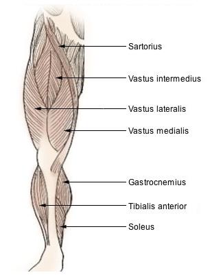
Vastus lateralis |
Greater trochanter, Intertrochantic line, Linea aspera of femur |
Patella via Quadriceps tendon, Tibial tuberosity via patellar ligament |
Knee:Extension, Stabilisation |
|
Vastus intermedius |
Lateral anterior femur (upper two-thirds) |
Patellar ligament |
Knee:Extension |
|
Vastus medialis |
Medial femur, from trochanters to lower quarter |
Patellar ligament |
Knee:Extension |
|
Rectus femoris |
Anterior inferior iliac spine, Iliac portion of acetabulum |
Patellar ligament |
Knee:Extension Hip: Flexion |
|
Sartorius |
Superior to anterior superior iliac spine |
Pes anserinus of the tibia |
Knee: Flexion Hip: Flexion |
The vastus lateralis, vastus intermedius, vastus medialis and rectus femoris all make up a the quadriceps femoris.
Adductor muscles of the thigh
|
Muscle |
Origin |
Insertion |
Actions |
|
Gracilis |
Ischiopubic ramus |
Pes anserinus |
Hip, Knee:Adduction, Flexion |
|
Adductor brevis |
Superior and inferior pubic rami |
Lesser trochanter, Linea aspera |
Hip: Adduction |
|
Adductor longus |
Pubis (below pubic crest) |
Middle third of linea aspera |
Hip: Adduction, Flexion |
|
Adductor magnus |
Ischiopubic ramus, Ischial tuberosity |
Femur |
Hip: Adduction, Extension |
|
Pectineus |
Superior pubic ramus |
Lesser trochanter, Linea aspera |
Hip: Adduction, Flexion, Medial rotation |
|
Obturator externus |
Medial to obturator foramen, Obturator membrane |
Greater trochanter |
Hip: Adduction, Lateral rotation |
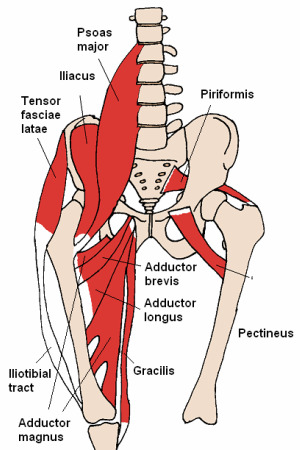
Image courtesy of commons.wikimedia.org/wiki/File:Anterior_Hip_Muscles_2.PNG under Creative Commons Attribution ShareAlike 3.0
Muscles of the buttock
|
Muscle |
Origin |
Insertion |
Actions |
|
Gluteus maximus |
Ilium, Sacrum, Lumbar fascia, Sacrotuberous ligament |
Lateral linea aspera, Iliotibial tract |
Hip: Extension, Abduction, Lateral rotation Knee: Supports extension |
|
Gluteus medius |
Ilium |
Greater trochanter |
Hip: Abduction, Medial rotation (when hip extended), Lateral rotation (when hip flexed) Support: When standing on one leg |
|
Gluteus minimus |
Ilium (below gluteus medius) |
Greater trochanter |
Hip: Abduction, Medial rotation (when hip extended), Lateral rotation (when hip flexed) Support: When standing on one leg |
|
Piriformis |
Anterior sacrum |
Greater trochanter |
Hip: Lateral rotation |
|
Quadratus femoris |
Ischial tuberosity |
Intertrochantic crest |
Hip: Lateral rotation, Adduction Stabilises:Femoral head |
Posterior muscles of the thigh
|
Muscle |
Origin |
Insertion |
Actions |
|
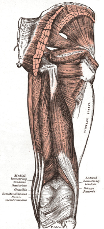
Biceps femoris |
Long head:Ischial tuberosity Short head:Linea aspera |
Head of fibula, Lateral condyle of tibia |
Knee: Flexion, Lateral rotation (when flexed) |
|
Semi-tendinosus |
Ischial tuberosity |
Pes anserinus |
Knee: Flexion, Medial rotation Hip: Extension |
|
Semi-membranosus |
Ischial tuberosity tendon |
Medial condyle of tibia |
Knee: Flexion, Medial rotation Hip: Extension |
These muscles all make up the hamstring.
Anterior and peroneal muscles of the leg
|
Muscle |
Origin |
Insertion |
Actions |
|
Tibialis anterior |
Lateral condyle of tibia, Upper two-thirds of lateral tibia |
Medial cuneiform, 1st metatarsal |
Foot:Dorsiflexion, Inversion |
|
Extensor digitorum longus |
Lateral condyle of tibia, Upper three-quarters of fibula |
Middle and distal phalanges of toes |
|