
Nerves and Blood Vessels
Nerves of the Leg
Nerves innervate muscles and skin. The nerves of the leg all originate from the lumbar plexus and sacral plexus. This group includes the sciatic nerve, femoral nerve, obturator nerve, tibial nerve and common fibular nerve (which splits into the superficial fibular nerve and deep fibular nerve).
Sciatic Nerve
The sciatic nerve is the longest, widest nerve in the body. It is derived from the 4th lumbar nerve and 3rd sacral nerve. It gives off muscular and articular branches.
The articular branches supply the hip joint and the skin of the leg.
The muscular branch splits into the tibial nerve and common fibular nerve which innervate all the muscles of the lower leg and some muscles of the foot (see below).
Femoral Nerve
The femoral nerve originates from lumbar nerves 2, 3 and 4 and is the largest nerve branching from the lumbar plexus. Shortly after crossing the femoral triangle, the nerve splits into several branches; some deep, some superficial.
The superficial branches are sometimes known as the anterior division. This includes both cutaneous branches and muscular branches.
The cutaneous branches are the intermediate and medial cutaneous nerves. The intermediate cutaneous nerve innervates the skin of the leg down to the knee. It splits into several smaller branches which communicate with a branch of the genitofemoral nerve at the proximal end of the thigh and also form the patellar plexus with the medial cutaneous nerve and branches of the saphenous at the distal end of the thigh. The medial cutaneous nerve gives off a branch which forms the subsartorial plexus with the obturator and saphenous nerves. The branches also innervate the skin on the medial side of the upper leg and communicate with the saphenous nerve.
The muscular branches include a branch to the sartorius and a branch to the pectineus.
The deep branches are sometimes known as the posterior division. This division includes the saphenous nerve and articular and muscular branches.
The saphenous nerve has several branches. One travels down the leg as far as the foot, where it communicates with the superficial fibular nerve. The nerve also gives off other branches which innervate the skin at the knee and on the front and medial sides of the lower leg. Some branches also communicate with other nerves including the cutaneous nerves, obturator nerve and subsartorial plexus.
The muscular branches of the posterior division supply the 4 muscles of the quadriceps femoris; the rectus femoris, vastus lateralis, vastus medialis and vastus intermedius.
The 3 articular branches supply the knee joint. One of these also supplies the synovial membrane.

Obturator Nerve
This nerve arises from the second, third and fourth lumbar nerves and enters the leg through the obturator canal, before splitting into anterior and posterior branches.
The anterior division provides innervation of the skin of the medial thigh, hip joint, adductor longis, adductor brevis and gracilis.
The posterior division supplies fewer areas, which include the knee joint and adductor magnus.
Tibial Nerve
The tibial nerve is a division of the sciatic nerve. It innervates the skin of the heel and many muscles of the lower leg. These include the gastrocnemius, soleus, popliteus, plantaris, flexor digitorum longus, flexor hallucis longus and tibialis posterior. The nerve terminates at the ankle, where it splits to the medial and lateral plantar nerves which supply the foot.
Common Fibular Nerve
This nerve is also known as the common peroneal nerve. It originates from the sciatic nerve and supplies the skin of the posterior, lateral surface of the leg. At the neck of the fibular bone, the nerve splits to give the superficial fibular nerve and the deep fibular nerve.
The superficial fibular nerve innervates the fibularis longus and fibularis brevis. It also supplies part of the skin of the foot and lower leg.
The deep fibular nerve lies further beneath the surface than the superficial fibular nerve. It innervates the tibialis anterior, extensor digitorum longus, fibularis tertius, extensor hallucis longus, extensor digitorum brevis, extensor hallucis brevis and ankle joint.
Blood Vessels of the Leg
The blood vessels supply the muscles and skin. Arteries carry oxygenated blood to the muscles, whereas veins transport deoxygenated blood away from the muscles.
Arteries
The femoral artery is found in the thigh and gives off several branches to supply the muscles in this area. These branches are the superficial external pudendal, deep external pudendal, superficial epigastric, superficial circumflex iliac, profunda femoris and descending genicular arteries . The femoral artery carries on proximally from the external iliac artery and becomes the popliteal artery distally.
The popliteal artery begins at the knee, where it is a continuation of the femoral artery. It supplies blood to some muscles of the thigh and lower leg, as well as supplying the knee joint itself. This artery then becomes 2 seperate arteries - the posterior and anterior tibial arteries.
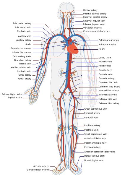
The anterior tibial artery supplies the anterior muscles of the lower limb. The dorsal side of the foot is also supplied by this artery. At the distal end of the artery it becomes the dorsal pedis artery, which supplys parts of the foot.
The posterior tibial artery takes blood to the posterior muscles of the lower limb and some areas of the foot. It has a branch known as the fibular artery. At the end of the posterior tibial artery it splits to become the medial plantar artery and the lateral plantar artery.
Veins
There are 2 major groups of veins; deep veins and superficial veins.
Deep veins generally follow the route of the arteries.
The femoral vein runs along the thigh next to the femoral artery. It anastomoses with the external iliac vein proximally and the popliteal vein distally. It has several branches. These are the profunda femoris vein and the medial and lateral circumflex femoral veins.
The popliteal vein is found running along the back of the knee, next to the popliteal artery. This vein then splits to become the anterior and posterior tibial veins.
The anterior tibial vein is found in the lower leg and runs alongside the anterior tibial artey. It gives rise to the dorsal vein in the foot.
The posterior tibial vein is also found in the lower leg and runs alongside the posterior tibial artery. It gives rise to several branches including the medial and lateral plantar veins (which meet at the deep plantar venous arch), the plantar digital veins and the plantar metatarsal veins.
Superficial veins tend to travel with the route of the nerves.
The great saphenous vein runs along the whole length of the leg from thigh to ankle. Here, it becomes the medial marginal vein. The small saphenous vein is a branch of the popliteal vein and runs lateral to the great saphenous vein in the lower leg. At the ankle it becomes the lateral marginal vein. The medial and lateral marginal veins meet at the dorsal venous arch. At this arch they give off branches known as the digital veins and metatarsal veins.
NOTE - there are small branches between superficial and deep veins to allow communication between them.